
|
For details scroll the page down
|
|
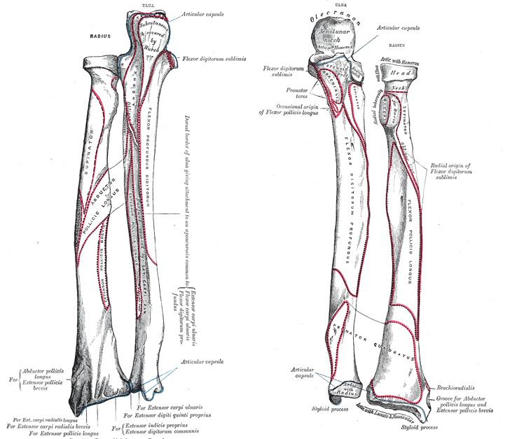
The Radial Bone
Radius is the lateral bone of the forearem being shorter than the Ulna. Proximally it articulates with the capitulum of the Humerus, distally with the carpus and with the Ulna medially.
It has got and upper end, a lower end and a shaft. Allthough both the ends are expanded, but the lower end is more wider than the upper end. The shaft increases in breath rapidly towards the lower end.
Side determination:
Small head is situated proximally with the radial tuberocity laying medially.
Large lower end is placed distally.
Smooth concave articular surface of the distal end faces inferiorly with pointed styloid process directed laterally.
Sharp interossius (medial) border facing medially.
Anterior surface of the shaft is concave, the lateral surface is convex while the posterior surface is concave at it's upper part and flat in the lower part.
Dorsal tubercle of listre must face posteriorly.
Upper end of Radius:
includes 1-head, 2- neck and 3-radial tuberosity.
1- Head: is cylindrical in outline and disc-shaped. It is concave & cup-shaped in it's upper surface for articulation with the capitulum of the Humerus.this concave surface is covered by hyaline cartilage in fresh bone. The articular circumference of the head is smooth and narrow rim like.
this circumference is deeper in medial aspect (where it articulate with radial notch of ulna), while lateral side of this rim is narrow being covered with annular ligament of the superior radio-ulnar union(joint).
2-Neck: is constricted cylindrical part of the upper end below the head. It is overhung (espescially on lateral side) by the head.
3-Tuberosity(Radial tuberosity): The neck is limited below by the radial tuberosity which is a conical projection placed distally to the medial part of the neck. It projects anterio-medially and separated the upper end of the radius from the radial shaft. It's posterior part is roughened but anteriorly it is smooth.
Shaft of the radius:
The shaft is generally curved with the convexity directed laterally. In transverse section shaft is triangular. The shaft has 3 borders and 3 surfaces.
Borders of the radial shaft.
Interossius(medial) border is the sharpest of the 3 borders and commences just below the radial tuberosity and is sharply defined in the middle 1/3rd and ends bellow by dividing into two ridges which extend upto the anterior and posterior margins of the ulnar notch of the radius. This terminal division of the interossius border inclosed a triangular area above the ulnar notch of radius. This triangular area is also an additional medial surface of the lower end. Lower 3/4 of this border gives attachment to the interossius membrane, which is distally attached to the posterior ridge only. This membrane connect the interossius borders of the ulna and radius forming the middle radio-ulnar union(joint).
Anterior border starts just below the anteriolateral aspect of the radial tuberosity running downwards with a lateral inclination from the tuberosity. This lateral curvature of the anterior border proximally is called "anterior oblique line". The anterior border can be traced to the ridge between the anterior and lateral surfaces of the lower end of radius.
Posterior border is the mirror image of the anterior border starting from the posterioinferior part of the radial tuberosity and runs downwards with lateral inclination from the radial tuberosity forming the "posterior oblique line" in the same way as the anterior border but couldn't be seem similarly in every bone because it is usually ill-defined. The middle part of the posterior border is distinctly defined and lower 1/3rd of the it is ill-defined where it ends at the dorsal tubercle of listre on the back of lower end.
Surfaces of the radial shaft:
Anterior surface lies between the anterior and the interossius borders of the shaft. It is slightly concave from side to side and curves forewards at lower end. A little above it's middle is situated it's nutrient foramen directed upwards towards the elbo joint.
Posterior surface lies between the interossius and the posterior borders. It is concave above and flattened to some extent below.
Lateral surface lies between the posterior and anterior borders and is gently convex in all directions. It about it's middle has an oval impression called pronator ridge or tubercle for the insertion of the pronator teres muscle. But below this impression the lateral surface is smooth.
Lower end of the radius:
Lower end is the widest part of the radius and is four sided in it's transverse section.
The lower end has five surfaces explained below.
1-Anterior surface is marked by a thick prominent ridge.
2-Posterior or dorsal surface is convex and is marked by Dorsal tubercle of listre mear it's middle. This tubercle is actually the terminal part of the posterior border of the radial shaft and divides the posterior surface of the lower end into medial and lateral parts. The tubercle is limited medially by a narrow oblique groove and a similar wider and undivided groove medially situated to the previous oblique groove.
A wider shallow groove lies lateral to the tubercle of listre and is divided into two parts by a very faint vertical ridge.
3- Lateral surface is rough and projects downwards beyond the rest of the bone to form the radial styloid process.
4-Medial surface: It is distally occupied by the ulnar notch of the radius for the ulnar head and is articular. The lower margin of this notch gives attachment ot the articular disc of the inferior radio-ulnar joint. Above the ulnar notch the medial surface of the lower end lies between the two bifurcations of the interossius border of the radial shaft and is roughened.
5-Inferior articular or carpal surface: this surface of the lower end is triangular in outline and is smooth. It takes part in the formation of the wrist joint. This surface is divided into a quadrangula medial and a triangular lateral areas by a faint ridge. The latera triangular area covers the medial side of styloid process and is for articularion with the scaphoid carpal bone and the medial quadrangular surface is for the articulation with the lunate carpal bone.
Attachments to the Radius:
The narrow articular circumference of the head and the adjacent parts of the neck are covered by the annular ligament of the proximal radio-ulnar union(joint).
The biceps tendon is inserted to the rough osterior part of the radial tuberosity, but the smooth anterior part of the radial tuberosity is separated from the biceps tendon by a bursa.
A little below the radial tuberosity the oblique cord of the middle radio-ulnar union(joint) is attached. This cord extends from ulnar tuberosity fo ulna to the radial tuberosity of radius.
Supinator muscle is inserted to the posterio-lateral aspect of the neck and upper part of the lateral surface of the shaft between the two oblique lines upto pronator ridge or tuberosity.
Pronator teres is inserted to the middle and the most prominent part of the lateral convexity of the lateral surface of the shaft at the pronator ridge or tuberosity.
The radial head of the flexor digitorum superfascialis arises form the anterior oblique line on the anterior surface.
Flexor pollicis longus takes origen from the upper 2/3rd of the anterior surface of the shaft.
Pronator quadratus is inserted to the lower 1/3rd of the anterior surface while it's deepest fibres are inserted on the medial side of the lower end above the ulnar notch and anterior to the posterior ridge fo the interossius border.
On the posterior surface below the posterior oblique line is the oblique origen of the abductor pollicis longus. This origen extends accros the interossius membrane upto ulna.
Below the above attachment the extensor pollicis brevis arises obliquely from a slightly hollow area and the adjacent part of the interossius membrane at the posterior aspect. The lower part of the posterior surface is bare and is covered by the above extensor tendons.
Attachments to the lower end:
Lateral surface of the lower end just above the styloid process recieves the insertion of the brachio-radialis muscle.
A narrow grooce medial to the dorsal tubercle of listre lodges the tendon of the extensor pollicis longus.
Medial to the above mentioned groove, the wider undivided groove lodges the tendons of extensor digitorum and extensor indicis.
Lateral to the dorsal tubercle of listre a wide shallow groove contains tendons of the extensor carpi radialis longus laterally and the the tendon of extensor carpi radialis brevis medially.
Tip of styloid process gives attachment to the radial collateral ligament of the wrist joint.
|
|
The Ulnar Bone
It is the longer and medial bone of the forearm (antebrachium). It is parallel to the radius during supine position of forearm. It articulates with the humerous above , with articular disc at wrist joint below , and laterally (above and below) with the radius at the superior and inferior radio-ulnar joints respectively.
Ulna tapers in reversed way to the radius. It is massive above and small at it's distal extreamity. It has got a shaft and two ends i.e. proximal and distal ends respectively.
Upper end of Ulna:
Upper end has got two substantial processes which are as follows:
Olecranon process
Coronoid process
and two articular surfaces which are as follows:
trochlear artculating surface
radial articular surface
Processes:
Olecranon process: It is the proximal extension of the shaft. It is bent forewards and forms a prominent beak which projects into the olecranon fossa of the humerus when forearm is extended.
Olecranon process has got 5 surfaces which are as follows:
Superior surface or upper surface which is rough behind for the insertion of triceps muscle and is smooth in front for a bursa that lies between the triceps tendon and the capsule of the elbo joint.
Posterior surface which is subcutanous and is generally ovoid or triangular in outline. It is smooth and lies between two oblique lines that meet bellow and continue downwards as posterior border of the shaft.It is covered by a bursa.
A demarcating ridge called the upper border of the
posterior surface or sometimes called "point of
elbow" is present between the superior and posterior
surfaces of the olecranon that can be felt on or just
above the line joining the epicondyls of humerus. This
is an importand landmark to exclude the dislocation of
elbow joint.
Anterior or articular surface which is smooth and articular and forms the upper part of trochlear notch. It is separated from the lower part of trochlear notch by a transverse groove or ridge and a constriction of the two sides which causes it to look like an hour glass.
Medial surface which is slightly concave and is continous downwards with the medial surface of the shaft.
Lateral surface that continues downwards with the posterior surface of the shaft and gives insertion to anconeus muscle.
Coronoid process: It is a bracket like projection from the upper end of the shaft just below the olecranon process. It fits into the coronoid fossa of the humerus.
Coronoid process has 4 surfaces that are explained as follows:
Superior surface that forms the lower part of the trochlear notch and is therefore smooth and articular.
Anterior surface which is triangular and bears on it's lower part the rough ulnal tuberosity.
Lateral surface which is somewhat triangular and uneven. The upper part of the lateral surface is marked by the radial notch for the radial head. This radial notch is separated from trochlear notch by a smoth ridge.
The lower part of the lateral surface is deepened to
accommodate the radial tuberosity of radius. This area
is limited behind by a ridge "called supinator crest".
Medial surface which is small and triangular area. At the anterior angle of the base of this triangular area is present a small but prominent tubercle which is called the "sublime tubercle".
Trochlear Notch is an Hour glass shaped articular area. It articulates with the trochlea of humerus. It is formed by the anterior surface of olecranon and the superior surface of coronoid process. This notch is constricted at the junction of the two areas which may be demarkated by a transverse ridge or groove into upper and lower part of the notch. It is also divided into a medial and a lateral part by a longitudenal smooth ridge that corresponds to the groove of the trochlea of the humerus. The trochlear notch as a whole is convex from side to side and is concave from above downwards.
Lower end of Ulna:
The distal or lower end of ulna is slightly expanded as compared to the upper end. It consists of following parts;
Rounded Head which presents a convex articular surface anteriolaterally for articulation with ulnar notch of radius. The convex articular surface is separated from styloid process by a triangular grooved area that gives attachment to the articular disc of the wrist joint. This disc separates ulna from triqueteral carpal bone. Inferiorly a crescentric area (below the anteriorly convex articular surface) is in contact with this triangular articular disc of wrist joint.
Styloid process is a small and conical projection from the posteriomedial side of the lower end. Proximally it continues with the posterior border of shaft. Posteriorly between the convex articular surface and styloid process there is a groove for tendon of the extensor carpi ulnaris muscle.
Shaft of Ulna:
The shaft is angled laterally from the line of trochlear notch. It is curved laterally in such a way that it's convexity is directed medially forming a carring angle with the arm. The shaft is triangular in it's transverse section in it's upper 3/4th but is almost cyindrical in it's lower 1/4th.
the shaft has 3 borders and 3 surfaces described bellow:
Borders of ulnar shaft:
Lateral or interossius border is sharp and well defined in upper 3/4th while is illdefined and indistinct in it's lower 1/4th and may be traced to latreral side of the head of ulna. Above it is continued by two lines or ridges that pass to margins of radial notch while in between these two lines a somewhat triangular depressed area called supinator fossa is present. This fossa lodges the tuberocity of radius during pronation. Regarding these lines of supinator fossa the posterior line or ridge is called supinator crest which ends superiorly at a tubercle called supinator tubercle just below this radial notch.
Anterior border is rounded and thick. Above it starts medially to ulnar tuberocity and also blends with the anterior end of medial margin of coronoid process. It passes downwards and somewhat anteriomedially and joins the medial side of base of styloid process.
Posterior border is subcutanous in it's whole extent and begins at the apex of the posterior subcutanous triangular surface of the olecranon process. From here it curves slightly laterally and then medially finally ending at the base of the conical styloid process.
Surfaces of ulna:
Anterior surface that lies between the interossius and the anterior border is concave in it's upper 3/4th. A little above the middle of this surface is a nutrient foramen that is directed upwards and transmitsthe nutrient branch of the anterior interossius artery. The lower 1/4th of this surface is crossed by an oblique ridge that is called pronator ridge that runs from interossius to the anterior border.
Medial surface that lies between the anterior and the posterior borders of the shaft of ulna is smooth throughout it's extent and is subcutanous in it's lower part.
Posterior surface that lies between the posterior and the interossius borders of the shaft looks slightly laterally. This surface is divided into 3 parts by two lines or ridges which are respectively called oblique and vertical ridge that are explained bellow:
Oblique line marks the posterior surface proximally and runs
upwards and laterally from the junction of the upper and the middle
1/3th of the posterior border and joins the posterior margin of the
radial notch at supinator tubercle.
Vertical ridge: The region below the above mentioned
oblique line is subdivided into a larger medial and a narrow lateral and
roughened part by a vertical ridge. This vertical ridge is prominent in
it's upper 3/4th but is usually less clear below.
The narrow lateral part of the posterior surface is subdivided into 3 areas by two oblique lines running from this vertical line to the interossius border. The upper area gives attachment to the abductor pollicis loingus, the middle area gives attachment to the extensor pollicis longus and the lower area gives attachment to the exensor indicis.
Side determination of the Ulna:
Proximal end is more expanded than the lower end.
Trochlear notch is facing anteriorly.
The interossius border is sharp and creast like facing laterally.
Radial notch is facing laterally.
Ossification of the ulna:
The shaft and most of the upper end ossifies from one primary centre that appears during the 8th week of the intra-uterine life.
A secondary centre appears for the superior part of the olecranon process during the 10th year. It forms a scale like epiphysis and that joins the rest of the bone at about 16th year.
Another secondary centre appears for the lower end about 5th year and ossifies by the 18th year and thus is the growing end of the ulna.
Attachments to ulna:
I Olecranon process has the following attachments:
Upper surface or superior surface of the olecranon process at it's anterior 1/3rd gives attachment to the capsule of the elbo joint, while it's posterior 2/3rd is roughened for the attachment of the tendon of triceps muscle and between these two attachments is present a bursa.
Medial surface of the olecranon process at it's upper part is marked by a rough elevation as a site of the attachment of the posterior and the oblique bands of the ulnar collateral ligament of the elbo joint and the ulnar head of the flexor carpi ulnaris muscle. The lower boundry of the medial surface of the olecranon is smooth and gives attachment to the uppermost fibers of the flexor digitorum profundus.
Lateral surface of the olecranon process and the adjoining posterior surface of the shaft above the oblique line gives insertion to the anconeus muscle.
Posterior surface of the olecranon process is triangular and featureless and separated from the skin by a bursa called the "olecranon bursa". The sides of this triangular surface gives attachment to the deep fascia of the forearm called "antebrachial fascia".
II Coronoid process has following attachments:
Anterior surface including the tuberocity of the ulna gives insertion to the brachialis muscle. The medial border of the anterior surface of the coronoid process is sharp. A rough and small sublime tubercle is situated at it's upper end for the attachment of the oblique and the anterior band of the ulnar collateral ligament of the elbo joint as well as to the origen of the ulnar head of the flexor digitorum superfascialis muscle (previously called flexor digitorum sublimus). Bellow the sublime tubercle , the medial margin of this anterior surface gives origen to the ulnar head of the pronator teres muscle. The ulnar head of the flexor policis longus may arise from the medial or more rarely from the lateral border of the anterior surface.
Medial surface of coronoid process is concave and gives origen to the flexor digitorum profundus.
Lateral surface of the coronoid process: The anterior border of the radial notch gives attachment to the annular ligament of the superior radio-ulnar joint. The supinator fossa below the radial notch is limited posteriorly by supinator crest. Supinator muscle arrises from supinator tubercle, supinator crest and the whole of the supinator fossa (in front of the supinator crest).
III Attachments to the shaft of the ulna:
Posterior border is subcutanous and gives attachment to the deep fascia of the forearm or the antebrachial fascia. This fascia attached to posterior border gives an additional origen in the upper 3/5th to the brachialis muscle and from the middle 1/3rd an additional origen to flexor carpi ulnaris muscle. So both of these muscles are connected to posterior border by this deep fascia.
Interossius border except it's upper 1/4th gives attachment to the interossius membrane.
Anterior border of the shaft at upper 3/4th gives origen to the flexor digitorum profundus.
Surfaces of the shaft:
Anterior surface at it's upper 3/4th gives origen to the flexor digitorum profundus in the same way as the medial surface and the anterior border of the shaft. It's lower 1/4th is marked by pronator ridge for the origen of the pronator quadratus muscle.
Posterior surface: Area above the oblique line including the lateral surface of the olecranon process and the coronoid process gives insertion to the anconeus muscle.
The area lateral to the vertical ridge is subdivided int three areas that pass from the vertical line to the interossius border. The upper area gives origen to the abductor policis longus muscle, the middle area gives origen to the extensor policis longus muscle and the lower area gives origen to the extensor indices muscle.
The medial part to the vertical line is devoid of muscles attachments but is covered or overlaped by the extensor carpi ulnaris muscle.
The tip of styloid process of the ulna gives attachment to the ulnar collateral ligament of the wrist joint.
Upper 3/4th of the medial surface gives origen to the flexor digitorum profundus muscle, while rest of the surface is covered by the medial muscles of the forearm.
|
|
|
|
|
|
|
|
|
|
|